
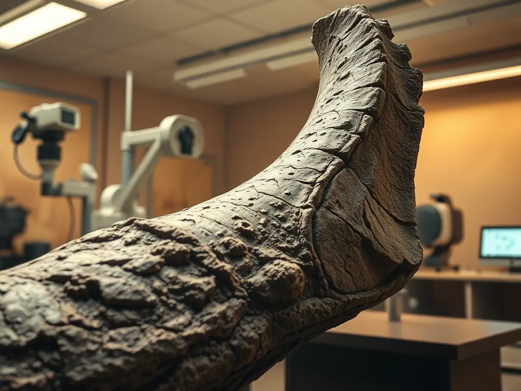
Diggers from the Royal Saskatchewan Museum found the skeleton of the tyrannosaur Scotty on August 16, 1991, in the Frenchman River valley in Saskatchewan, Canada. Thirty-five years later, one of its ribs has turned out to hold a web of fossilized blood vessels — left over from the animal's attempt to heal a broken bone 66 million years ago. Nothing like it has ever been seen in another fossil. Scientists spotted it without cutting the bone, using neutron and synchrotron imaging, methods like X-rays but far better at picking up soft, organic remains buried inside dense rock.
A physics student's find in 2020
In 2020, Jerit Mychak, then a physics student at the University of Regina, spotted traces of blood vessels in Scotty's rib while checking it with a micro-CT scanner, an X-ray method that leaves the sample undamaged. He kept studying the fragment with his supervisor, professor Mauricio Barbi, and colleague Marcela Berg, using synchrotron radiation at the Canadian Light Source — a particle accelerator that produces very bright X-rays for detailed images — and microscopy to trace the healing down to cell level. In April 2026, the team brought the rib to Oak Ridge National Laboratory in the US, where two neutron instruments, MARS and VENUS, confirmed the earlier findings and built a full 3D image of the large bone without a single cut.
MARS uses cold neutrons, which pick up soft tissue especially well, while VENUS works with higher-energy neutrons and builds the complete 3D picture of the rib. Hassina Bilheux leads the VENUS instrument. Scientists rank the Frenchman River valley, where Scotty was dug up, as the richest dinosaur fossil site in North America. "It's like winning the lottery," Barbi says of finding the blood vessels preserved, while Mychak calls each fossil a small snapshot of a bygone time.
Goethite, pyrite and two layers of mineralization
The synchrotron images show the vessel network at a resolution of nearly 18 micrometres — sharp enough for scientists to trace how it branches from healthy bone tissue into the callus, the new tissue the body builds around a break as it knits it back together. Individual vessels measure between 100 and 500 micrometres across, with some reaching up to 1 millimetre. The team also used X-ray fluorescence, which shows exactly where iron sits in the bone, and a method called XANES, which shows what chemical form that iron takes.
Under the microscope, the vessels show two separate layers of mineralization — a structure typical of blood vessels that grow during healing, not of minerals that simply seeped into the bone by chance. Chemically, the vessels are preserved mostly as goethite, an iron oxide mineral that makes up about 96% of the finds, with traces of pyrite. After the rib broke, iron-rich blood flooded the wound, and Scotty's body began building new vessels — a process called angiogenesis — to feed the site with nutrients and speed up healing. Soft tissue almost never survives fossilization, because bacteria and decay destroy it long before minerals can replace the bone, which is why a find like this is so rare in paleontology. The healing was never finished: the animal died with the rib still not fully mended, and the body ended up in a salt marsh, where conditions slowed decay just enough for the fragile vessel network to fossilize instead of vanishing.
The skeleton at the T. rex Discovery Centre in Eastend
Scotty, catalogued at the museum as RSM P2523.8, measures between 12 and 13 metres long and stands about 4 metres tall at the hip. At an estimated weight of nearly 8.9 tonnes, it is the heaviest known Tyrannosaurus rex specimen. Early estimates put its age at death around 30 years; later analysis lowered that to between 23 and 27 years, but Scotty still ranks among the oldest known tyrannosaurs. The skeleton is kept at the T. rex Discovery Centre, run by the Royal Saskatchewan Museum, in Eastend, and a cast has been on display at the museum in Regina since 2019.
The University of Regina team says that after the discovery in Scotty's rib, other fossils showing signs of injury or fracture are now natural candidates for neutron and synchrotron scans.

Comments (0)
Все още няма коментари.